Helpline 1800-120-4556
goalbertdiagnostics2018@gmail.com
A DIVISION OF GUT GI AND LIVER HOSPITAL

Gastroenterology Services

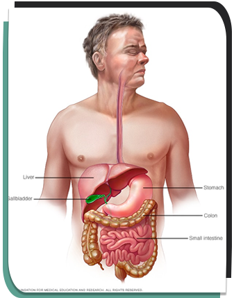
Gastroenterology involves the treatment and diagnosis of digestive system disorders that affect the oesophagus, stomach, small and large intestines, rectum, liver, gallbladder and pancreas. 
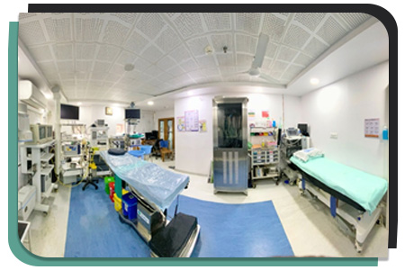
Most GI studies and procedures are performed on an outpatient basis in Gut Hospital state-of-the-art Endoscopy Suite. The Endoscopy Suite backed by senior gastroenterologist and experienced technician staff offers a welcoming and comfortable environment for patients.

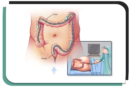
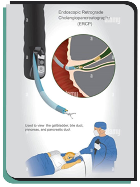
Our endoscopy room is equipped with an armamentarium of latest HD scopes exploring upper GIT (Gastro-scope), Lower GIT (Colono-scope) and Pancreaticobiliary tree (ERCP), Endoscopic Ultrasound (EUS) enabled with Histogram and Liver probe to analyse vascular details as well stiffness of tissue which is valuable in chronic liver disease as well as in cancers.

All standard protocol of infection control is followed for to patient care & prevent cross infection. High end Endo washer are used for cleaning scopes and UV light cabinets are used for their storage. Full time anesthetist are available to assist in all procedure and provide support to apprehensive patients.

Endoscopy

Endoscopy is the examination and inspection of the interior of body organs through an endoscope. An endoscope is a device using fibre optics and powerful lens systems to provide lighting and visualization of the interior of a body organs or cavity. It us inserted through are mouth and we can see duodenum. The portion of the endoscope inserted into the body may be rigid or flexible, depending upon the medical procedure. We require Endoscopy in following condition:

  • Persistent Heartburn
  • Nausea & Vomiting
  • Abdominal Pain
  • Difficulty Swallowing
  • Gastrointestinal Bleeding
  • Unexplained Weight Loss

Upper GI endoscopy can be used to identify many different diseases:

  • Gastroesophageal reflux disease
  • Ulcers
  • Cancer
  • Inflammation, or swelling
  • Precancerous abnormalities such as barrett’s esophagus
  • Celiac disease
  • Strictures or narrowing of the esophagus
  • Blockages

Colonoscopy

A colonoscopy is a medical procedure that involves using a long, flexible tube or scope to look for abnormalities, polyps, and growths in the rectum and colon (large intestine).The large intestine helps in the digestion and absorption of food and eliminating waste. The colon empties into the rectum, and the waste leaves the body through the anus. A colonoscopy is commonly used to evaluate gastrointestinal symptoms, such as bleeding, abdominal pain, rectal bleeding, chronic diarrhea and other intestinal problems.

Colonoscopy can help doctors diagnose the reasons for:

  • Unexplained changes in bowel habits
  • Abdominal pain
  • Bleeding from the anus
  • Unexplained weight loss

Colonoscopy is performed to detect inflamed tissue, ulcers, and any abnormal growth. This procedure helps your doctor to detect early signs of colorectal cancer. Your doctor can also take samples of abnormal tissues during colonoscopy for biopsy. Your doctor can also remove polyps and take biopsies using tiny tools passed through the scope.

Colonoscopy can be used to:

  • Remove polyps (polypectomy)
  • Dilate narrowed segments (stricture dilation) of large intestine and place metallic stents across them (colonic stenting)
  • Banding for haemorrhoids (piles banding)

FibroScan

A FibroScan® is used to assess the degree of fibrosis (scarring) in your liver. This is a relatively new technique designed to estimate the amount of scarring without having to do a liver biopsy. The technique is very similar to an ultrasound, but much quicker. A probe is held against the skin and generates a pulse sending a wave through the liver. The speed that the transmitted wave travels though the liver can be measured by the probe. The stiffness of the liver influences the speed of the wave, and in turn we can measure the “stiffness” of the liver. This measurement correlates with the degree of liver fibrosis which can then be estimated. So this procedure is used in patients who have been diagnosed with or are being monitored for hepatitis C (HCV), chronic hepatitis due to hepatitis B (HBV), a combination of hepatitis C and HIV, alcoholic liver disease, hepatic steatosis (fatty liver) non-alcoholic, chronic cholestatic diseases, liver cirrhosis, and monitoring for pre- and post-liver transplant patients.

There are several things that can harm the liver, including:

  • Infections that affect the liver – Inflammation of the liver (Hepatitis). Common causes are the viruses Hepatitis A, B, C.
  • Non-Alcoholic Fatty Liver Disease – Fatty liver disease affects approximately 20 percent of the population worldwide and is commonly seen in people with diabetes and obesity. People with fatty liver disease may progressively damage their liver to an extent that requires a liver transplant.
  • Abuse of alcohol – This is the most common cause of cirrhosis in the Western world and represents one of the ten most common causes of death.
  • Liver damage – If the liver sustains damage from any cause, normal liver tissue can become:
    • Fatty (steatosis)
    • Fibrous (fibrosis)
    • Scarred (cirrhosis)

ERCP

A procedure to diagnose possible disorders of the pancreas and bile duct system. Bile is made in the liver and stored in the gallbladder until food is eaten. Bile is then discharged into the cystic bile duct and travels to the common bile duct. The pancreas produces digestive enzymes that flow into the common bile duct. Together, bile and the pancreatic digestive enzymes travel through the ampulla (the point at which the common bile duct meets the duodenum) and into the duodenum to help digestion. Common problem requiring ERCP are:

  • Blockages or stones in the bile ducts
  • Fluid leakage from the bile or pancreatic ducts
  • Blockages or narrowing of the pancreatic ducts
  • Tumors
  • Infection in the bile ducts

Cholangioscopy

Cholangioscopy allows direct visualization and subsequent therapeutic maneuvers of the biliary ductal system. With advances in endoscopic and imaging technology, cholangioscopy has become an important modality for the diagnosis of indeterminate biliary strictures and an essential therapeutic tool for difficult to remove biliary stones. Enhanced imaging and operability of the latest generation cholangioscopes have further expanded their clinical applications to include ductal tumor ablation, gallbladder drainage, access to difficult to reach branches of the biliary tree, and biliary foreign body manipulation. In this review, we discuss the technical evolution of cholangioscopy into the digital era and review the clinical evidence supporting its use in the diagnosis and therapy of biliary tract disease.

How Is Cholangioscopy Performed?

This endoscope marvel from BOSTON SCIENTIFIC is extreme useful tool for biliopancreatic diseases. Direct Visualization system is an intuitive platform that enables simple, single-operator, direct visualization cholangioscopy for detection and treatment of large stones and strictures throughout in the pancreatico-biliary system. By attaching the single-use Spy Scope Access and Delivery Catheter to a standard duodenoscope and inserting through the working channel

Spy Glass Direct Visualization System allows for:

  • Site-specific examination of the entire lumen
  • Improved diagnosis by visualization and Spy Bite Forceps biopsy
  • Treatment of large biliary stones

It is extremely useful modality for patient with:

  • Obstructive Jaundice
  • CBD Stones & intra hepatic stones
  • Cholangio Carcinoma & indeterminate biliary strictures
  • Removal of inward migrated biliary & pancreatic stents

Spy Glass DS II


(Direct Visualization System in ERCP)